www.magazine-industry-usa.com
02
'25
Written on Modified on
How Stratasys Differentiates Itself in Radiopaque 3D Printing
The pioneering Stratasys material lets US healthcare providers create ultra-realistic patient-specific 3D models with tunable X-ray visibility for enhanced medical training and device testing.
www.stratasys.com
Radiopaque 3D-printed models are increasingly used in CT-based device testing, imaging protocol optimization, clinician training, and the creation of anatomically realistic phantoms for research. Their value lies in replicating human tissue visibility on X-ray-based modalities, enabling controlled, repeatable, and highly standardized imaging environments.
A New Material Class for Imaging-Realistic Models
Stratasys has made its RadioMatrix™ material fully commercially available in the United States, expanding access beyond initial limited deployments. RadioMatrix is currently the only 3D printing material offering fully tunable, repeatable radiopacity, enabling users to precisely control how printed models appear on CT and other X-ray-based imaging.
This capability positions the material differently from conventional 3D printing polymers, which cannot mimic the complex radiodensity variations of human tissues. With RadioMatrix, healthcare providers, research institutions, and device manufacturers can produce patient-specific or experimental phantoms with radiographic behavior closely aligned to real anatomy.
Validation Through Industry Collaboration
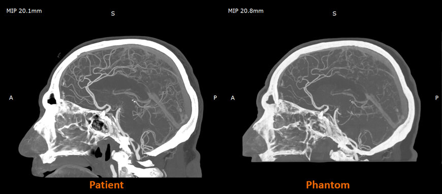
Research carried out with Siemens Healthineers has demonstrated that RadioMatrix-based phantoms can reproduce human tissue radiodensity with deviations as low as single Hounsfield units in key structures, including grey matter and venous anatomy. The partnership also showed that combining Stratasys Digital Anatomy™ technology with the radiopaque material enables models that preserve fine anatomical detail and pathological variations while remaining consistent across prints.
Compared with traditional phantom manufacturing and cadaver-based testing, this approach offers greater standardization and scalability. It also allows researchers and clinicians to iterate rapidly when developing new CT algorithms, calibrating imaging systems, or validating device behavior.
Early Clinical and Research Use Cases
Work in the UK with partners such as CPI and Beaumont Hospital is illustrating how radiographically realistic phantoms can raise the fidelity of imaging-based training. Cerebral angiography models printed with RadioMatrix are used to create controlled, repeatable practice environments—conditions that are difficult to achieve with cadavers or standard phantoms.
Impact on Imaging Education and Device Development
The broader U.S. availability of RadioMatrix marks a significant step in expanding access to radiographically accurate, customizable models. According to Stratasys, this advancement enables radiologists and device developers to transition from traditional phantom solutions to scalable, reproducible 3D-printed alternatives.
By providing anatomically realistic and radiographically precise phantoms, the technology is expected to accelerate CT protocol optimization, support the development of advanced imaging algorithms, and improve training for interventional and diagnostic radiology.
www.stratasys.com